Search for:
Cutting edge: Raman directed cancer surgery

Successful cancer surgery depends crucially on accurate removal of all the cancerous material. This can be tricky in cases where the tumor has invaded healthy tissue with its crabby legs and pincers. Simply erring on the safe side and removing large amounts of surrounding tissue isn’t always a good solution because this can affect the functioning of adjacent tissue.
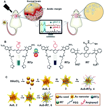
raman cancer cell molecular probe
Cancer cell molecular Raman probe (by Dept Neurosurgery, Fudan University from Chemical Science Journal)

Avoiding unnecessary damage to healthy tissue surrounding a tumor in the brain is particularly important. It is also particularly difficult because the human brain has upwards of 85 billion nerve cells and something like a million billion interconnections. There are however some tell-tale signs to help guide surgery. It has been known for some years that tumor cells are frequently more acidic than normal healthy cells and this month a group from the Fudan University in Shanghai reported a novel way to guide the removal of brain tumor cells based on the fact they are more acidic than normal cells.

Writing in the journal Chemical Science Ying Mao, Xiao Yong Zhang and Cong Li describe how they designed a molecular probe that selectively reveals differences in acidity and used it to guide the removal of rat brain tumors. Importantly, surgery guided by the use of the molecular probe during the operation gave more complete removal of the tumor compared to standard pre-operative MRI imaging and with less removal of healthy tissue compared to use of a control dye probe during surgery.

They synthesised special dye molecules and attached them to gold nanoparticles, which made them easy to detect using a technique known as surface-enhanced resonance Raman scattering (SERRS). Normally Raman scattering gives scientists a very weak signal but when attached to the gold particles the light emitted by the probe molecules increased millions of times.

The Fudan group call their new cancer probe AuS-IR7p. Au refers to the coupling to gold nanoparticles; IR7 refers to the fact the dye is excited with near infrared light (which penetrates tissue much better than shorter wavelengths); and the p stands for the fact that when the dye is in an acidic environment (protonated) it apparently changes its orientation on the gold nanoparticle which changes the relative intensity of two of the characteristic peaks in the Raman spectrum. This proved very useful because taking the ratio of the two peaks gave a measure of the acidity of the brain tissue that was independent of dye concentration and overall fluorescence intensity.
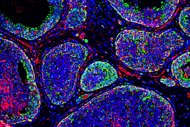
Conventional fluorescent dye staining to reveal acidic boundaries (in red) around tumor cells (by N. Rohani from MIT News / March 2019)

The neurosurgery project at Fudan University used a classic approach to innovation. While the acidic nature of cancer cells was well-known, it was only last year that the Koch Institute at MIT reported the detection of tumor cell boundaries using fluorescent dye. Putting together new information from an established area of need and novel solutions to problems using existing dye probes they have developed a potentially valuable innovation.

It remains to be seen if further studies confirm the advantages of Raman directed brain surgery but as a case study in innovation it makes fascinating reading.

The original Chemical Science journal article published by the Royal Society of Chemistry can be found here. Additional experimental details can be found here. Background on the MIT group research can be found here and the literature paper is published in Cancer Research.
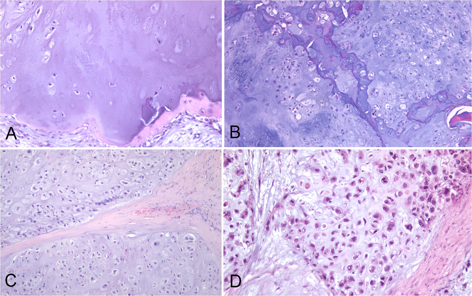
H+E stained tissue
Bone cancer diagnosis by Raman microscopy

Pathologists have an unenviable job. Armed with just their eyes, their experience and a pinky-coloured picture they must decide whether a patient requires life-saving surgery, has nothing to be concerned about or just needs to be kept an eye on.

h+e stained biopsy images
H+E stained tumor biopsies A) EC, B)CS:G1, C) CS:G2, D) CS:G3 (from Nature Scientific Reports)

No matter which part of your body might have started behaving badly, a biopsy is the most likely procedure to reveal a potential problem or give the reassuring all-clear. Once the biopsy sample has been taken it is preserved, thinly sliced and then typically stained with H+E (hematoxylin and eosin) stain. When an experienced pathologist looks at a microscope image of a good quality stained sample, they are extremely successful in classifying the tissue and providing an accurate diagnosis.

Even though H+E staining has been used for more than 100 years to reveal the microscopic structure of different tissues, there is no single protocol or set of chemical reagents to produce identical results time and time again. Each pathology lab technician will have their own preferred way of preparing stained biopsy samples. The introduction of digital imaging of the view down the microscope has made it easier and quicker for a pathologist to assess a particular biopsy sample but variability in staining persists and can make it challenging to classify different cell morphologies. Staining is cheap and widely used but can be time consuming and does not always give pathologists the quality of images they need.

For these reasons, researchers have been developing Raman microscopy as an alternative pathology tool. Dr Mario D’Acunto at the IBF-CNR, Istituto di Biofisica and colleagues at the University of Pisa recently reported a pilot study of patients with bone tumors. Publishing in Nature Scientific Reports, they describe how tumors can be classified by Raman microscopy without the need of stains or time-consuming sample preparation. Using statistical methods on Raman microscope images of biopsy samples, they obtained 90% sensitivity, 90% specificity and 90% accuracy in classifying tumor types. This is comparable with the performance of expert pathologists assessing the best quality stained images.

So how does Raman microscopy work and what are the statistical methods D’Acunto et al used? In Raman microscopy a laser is scanned over the surface of the biopsy sample and the light scattered by the sample gives a kind of fingerprint of the different molecules present at each point in the image. The fingerprint is known as its vibrational spectrum. Information about even subtle changes in the way molecules are joined together can be extracted from individual points in the image. It is also possible to use statistical methods to extract similarities and differences between the vibrational spectra of the whole Raman image. These similarities are known as Principle Components and by carefully comparing the relative amounts of different Principle Components of each type of biopsy, it was possible to classify different tumor types.

Accurate bone tumor classification based on biopsy is crucial. Benign EC (enchondroma) tumors are not concerning but CS (chondrosarcoma) tumors account for around 20% of malignant bone tumors and must be detected. Of increasing concern are CS1 (chondrosarcoma 1) which rarely metastasize (spread) and CS2 and CS3 which can metastasize in up to 70% of cases. Catching CS type tumors quickly is therefore important and correctly differentiating CS2 and CS3 from CS1 types is crucial so that effective treatment can be prioritised.

Raman microscopy therefore looks to have an important role to play in the future of pathology and, cancer diagnosis and treatment. Combined with statistical methods and new artificial intelligence tools, major advances in the 100 year old science of tissue imaging are knocking at the path lab door.

The whole journal article can be found at https://www.nature.com/articles/s41598-020-58848-0 . A practical introduction to H+E staining can be found on the Leica microscope website.