Search for:
Finding rare earth elements by hyperspectral imaging
Rare earth elements (15 lanthanides plus Scandium and Yttrium)

Imagine a world without smart phones, the latest medical imaging or great bass response from your earphones? Nope, nor can I. Worryingly, the continued availability of these essential 21st century pieces of tech is far from certain because they each rely on so-called Rare Earth Elements (REEs). REEs are not particularly rare but they are rarely found in the earth at sufficiently high concentrations to make it commercially viable to mine them.

The vast majority (>90%) of the world’s supply of REEs comes from China; the USA and Australia producing most of the remainder. With the fast growing demand for REEs and the equally fast growing economic Cold War between China and the West, supply is looking precarious.

Historical REE production 1958 – 2008 (courtesy US Geological Survey)

In 2016 a detailed report was published by the European Union EURARE project. European geological survey groups from Finland to Greece reported rare earth elements were widely present across Europe but there were just a few potentially viable REE mines. The figure below shows the EURARE distribution of ‘belts’ of REE geology. The next step would be to send a geologist with her rock hammer out into the field and then drill cores to find exactly where viable concentrations of rare earth minerals could be mined. But how to examine these areas of hundreds of square miles of land?

Belts of potential European REE geology (image courtesy of Elsevier Ore Geology Reviews)

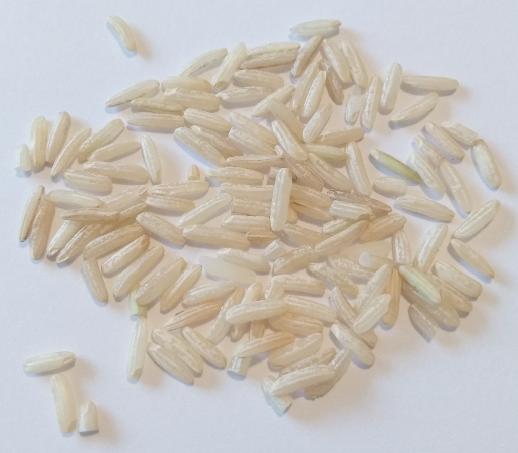
Researchers from the Helmholtz Institute in Freiberg in Germany believe they have found the answer. Rene Booysen and her colleagues reported this month that Unmanned Aerial Vehicles (UAVs) can be used to deploy hyperspectral imaging cameras to accurately locate REE deposits on the earth’s surface. Lanthanide minerals absorb light in very characteristic ways. Rather than absorbing broadly yellow light like a sapphire for example, the lanthanide REEs have very narrow absorption bands. Narrow bands are loved by spectroscopists because they can be much more easily picked out against backgrounds. Providentially, REEs tend to be found together and one lanthanide in particular, neodymium Nd (see the blog title image), absorbs light very strongly indeed in its narrow absorption bands.

Reflectance spectra from the surveyed area in Siilinjaervi Finland, showing narrow absorption peaks due largely to REE neodymium. Survey points contain increasing amounts of neodymium. (courtesy Nature Scientific Reports)

The Helmholtz Institute group cleverly exploited this fact by mounting a hyperspectral imaging camera on a drone and then rapidly covering 10,000 m2 areas of interest in Namibia and Finland. In addition to the hyperspectral camera on a copter drone, a small UAV plane was used for 3D imaging so that geometric corrections could be made to the images taken by the drone.

UAV plane (A) used to correct HSI images and copter drone (B) with HSI imaging camera (courtesy Nature Scientific Reports)

Areas in Finland and Namibia were pre-selected from geological regions rich in so-called carbonatites, rocks on the surface known to contain REE minerals.

Geological maps of Namibia (A) and Finland (B) showing locations of carbonatite rocks where REEs can often be found (courtesy Nature Scientific Reports)

Impressively, comparison of the HSI neodymium images with elemental analysis of rock samples from the survey sites in Namibia and Finland showed good agreement with concentrations of neodymium and other rare earth elements.

Further extensive surveying and mining in Europe will be required to establish what mineable reserves of essential REEs Europe holds. The team from Helmholtz Institute in Germany have nevertheless validated hyperspectral imaging as a major step towards securing the economic future of many key modern technologies.

Read the full Nature Scientific Report here.

The European EURARE report was published by Elsevier here.

More information on rare earth minerals and geology can be found at the American Geosciences Institute.

Hyperspectral imaging reveals rice quality and growing conditions
brown rice
smartphone photo of brown rice (copyright Corbeau Innovation)

Hyperspectral imaging (HSI) sounds like something Superman should have. And indeed it is. Hyperspectral imaging is increasingly used in art conservation, waste management, industrial quality control, medicine and agriculture. HSI not only gives an image of an object like the smartphone photo above, it also contains a detailed spectrum of every single pixel in the image.

Most images you find on Google or Instagram are either black and white, greyscale or colour. Colour information is typically restricted to how much red, green and blue light make up a particular point. This is because cells in the human eye measure only these three colours. Spectral imaging, on the other hand, records many different wavelengths of light. Hyperspectral imaging records a wide range of wavelengths with high resolution.

Whipping out your smartphone and taking a picture of a map, a set of instructions or a business card has replaced note taking. Clicking an image is faster, more reliable and durable than laboriously writing on scraps of paper. You can return to a saved image and rather than having to de-cypher poor writing or gaps in instructions, you can zoom into more detail than you realised was there in the first place. The same is true of hyperspectral imaging compared to conventional measuring and documenting.

Researchers at the US Department of Agriculture (USDA) Dale Bumpers National Rice Research Center in Arkansas have used these advantages of hyperspectral imaging to demonstrate a quick and reliable way to assess rice quality. Rice is the second most important food staple globally and some 3 billion people rely on rice as their main foodstuff. It is a truly massive commodity and assessing rice quality quickly has global importance.

The USDA team’s research demonstrated HSI was capable of: distinguishing different types of rice; telling which soil type the rice was grown in; quantitatively measuring the chalkiness of a particular type of rice. This final capability is possibly the most valuable because so-called chalk in rice is due to agglomeration of starch particles within the seed and it makes the grains break when they are milled. Fractured grains are useless for human consumption and identifying batches of rice with a high chalk content prior to milling saves money and improves efficiency.

Rice chalk prediction based on HSI and compared to actual SEM values
Comparison of rice images, % chalk and HSI (A); HSI predicted vs actual % chalk (B); SEM chalk content for comparison (C-E) (courtesy USDA/Nature Scientific Reports)

The methods used to uncover the hidden information in hyperspectral rice images are well-known to data scientists but less-so to agronomists. Dividing the spectra from the grain images into narrow wavelength windows, it was possible to find windows and combinations of window values that acted as components from which the spectrum of any pixel in the spectral image could be reconstructed. These Principle Components were then used to produce a calibration plot of HSI predicted %chalkiness (derived from Partial Least Squares/Discriminant Analysis PLS-DA) against true %chalkiness. The figure above shows a convincing linear relationship between the HSI parameter and the actual % chalk measured by SEM.

Hyperspectral rice images were also used to reveal which batches had been grown in Arkansas (AR09 & AR10) and which in Texas (TX08). Relative intensities of spectral windows are plotted in the figure below. PLS-DA analysis was used to make a classification of each of the three type of rice, TX08, AR09 and AR10. The red, green and blue rice batches can be clearly seen to cluster in distinct groups, with a small number of overlapping points.

PLS-DA analysis of hyperspectral rice images to discriminate environmental growth conditions
Differences in spectral window parameters from Texas rice (red) and Arkansas rice (blue and green) (A); discrimination of Texas rice (red) from Arkansas rice (blue and green) (C-E) (courtesy USDA/Nature Scientific Reports)

Finally the hyperspectral rice images were used to demonstrate discrimination of three sub-species of rice: IND (indica), TRJ (tropical japonica) and TEJ (temperate japonica). The figure below shows again the differences in the spectral windows used in PLS-DA analysis of the three batches of rice.

PLS-DA analysis of hyperspectral rice images to discriminate sub-species
Discrimination of TRJ, TEJ and IND rice types based on PLS-DA analysis of hyperspectral intensity values (courtesy USDA/Nature Scientific Reports)

The TRJ, TEJ and IND clusters are quite widely spread but clearly separated by analysis of the hyperspectral images.

A full account of the agronomy and the methods used in the study can be found at https://doi.org/10.1038/s41598-020-65999-7 in Nature Scientific Reports. Figures are used under Creative Commons with thanks to USDA and Nature. The full report also gives an interesting account of the genomics of the rice sub-species used and some consideration of Single Nucleotide Polymorphism (SNP) differences that may be seen in the hyperspectral phenotypes.