New blood test for Duchenne Muscular Dystrophy
Duchenne Muscular Dystrophy (DMD) affects almost exclusively boys and there are around 2,500 people with the disease in the UK. It develops rapidly from around age 4 and is a life-limiting condition, with muscle tissue gradually degrading until it becomes lifeless. Life expectancy is typically 26 years. As a rare disease, research into DMD is not widely funded but research is also held back by the lack of a quick, definitive blood test. Such a test would permit studies to start at an early stage and allow treatment to slow the progression of the disease to start earlier too.
This month, researchers at the University at Albany in New York reported development of a new blood test for DMD. Writing in Nature Scientific Reports, they validated the test in so-called mdx mice (which have the disease). Encouragingly, they gave a measurably different response to healthy control mice.
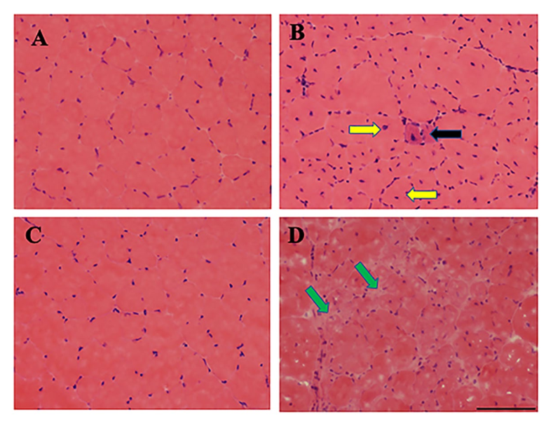
The usual way to diagnose DMD is with a muscle biopsy. Even with stained tissues, it can be difficult to tell the difference between healthy and diseased samples. White fatty regions can be fairly easily seen in 12 months old mice but at 3 months, differences are not so obvious.
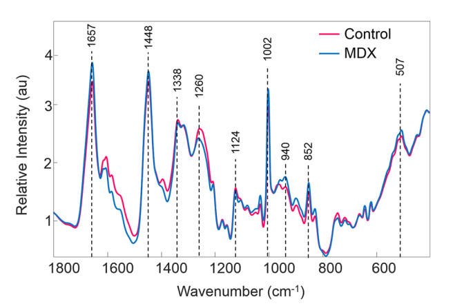
Principal Investigator Professor Bijan Dey and his team took blood samples from the diseased and healthy mice, separating the blood plasma from the red blood cells and other components in the blood. Drops of blood plasma were allowed to dry and then multiple points on the residues were analysed with a Raman microscope.
Laser Raman microscopy is a non-destructive analysis technique that gives a ‘finger-print’ of the vibrations of molecules in a sample under analysis. Large molecules have many vibrations and give complex Raman finger-prints, while small molecules have just a few and give simple finger-prints. Heavy molecules vibrate at low frequencies whereas light molecules vibrate at high frequencies. The finger-print is a spectrum of these Raman vibrations, from low frequencies to high frequencies.
The figure at the top of this blog shows the subtle differences the Albany team found between blood from the healthy mice group (in red) and the diseased mice group (in blue). Looks easy to imagine how measuring at just a few points in the spectrum would give a simple test doesn’t it?
Actually it involved quite a bit more work. The figure shows averages of all the blood samples used in the test and the differences between individual tests were not always obvious. To find a way around the variation in the data in each group the researchers used a statistical method known as PLS-DA (Partial Least Squares – Discriminant Analysis). This mathematical method allows just the parts of the finger-prints that change the most to be used to classify a blood sample as either diseased or normal. Half the blood samples were used to devise a PLS-DA classification model which was then applied to the remainder.
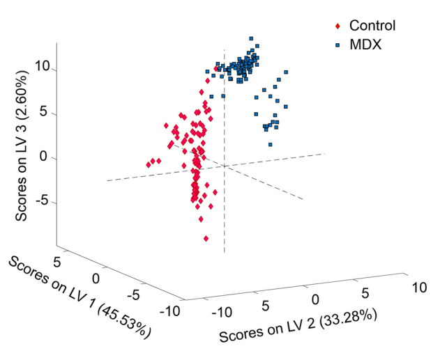
Plotting three discriminating variables (LV1, LV2 and LV3) derived from the PLS-DA model, the figure above shows how the two clusters of blood sample results are clearly separated.
There is still much to do to fully validate the new Raman blood test for DMD. mdx mice are a widely model for Duchenne Muscular Dystrophy but further work is necessary to extend these promising results to patients.
Further information about Duchenne Muscular Dystrophy can be found at the National Organization for Rare Disorders. The Nature Scientific Report can be downloaded here.