Search for:
malling centenary strawberry imaging
Smartphone spectral leaf imaging
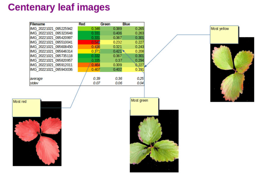
malling centenary strawberry imaging
Strawberry leaf images and Red Green Blue indices derived using OpenCV image analysis

All season we have been monitoring the health of our Strawberry Greenhouse crop. In addition to visual inspection with a loupe, digital leaf imaging has been a useful way to follow the development of the plants. Now that autumn has arrived, the leaves are showing the usual spectacular colour changes.

Chlorophyll green quickly gives way to yellowing and eventual reddening of the leaves. The yellow colour pigments are usually present in the leaves but as chlorophyll production is much less stimulated by sunshine, the yellow pigments can be seen. Red pigments are increasingly produced in leaves as the sugar concentration in the leaves increases. Leaf yellows are due to carotenoid pigments and leaf red colours are due to anthocyanin pigments.

Chlorophyll molecules absorb both red and blue light, leaving green visible light to be reflected by plant leaves, making them appear green. Carotenoid molecules  on the other hand absorb light in the blue end of the spectrum making leaves reflect and scatter yellow, green and red light. Anthocyanin molecules absorb blue and green light, making leaves reflect and scatter deeper red light.

leaf pigments spectra
Absorption spectra of chlorophyll a (blue), chlorophyll b (green), carotinoid type (orange), anthocyanin type (red) (Courtesy SPIE journals Creative Commons and Universidad de Guadalajara, Mexico)

Spectrometers and hyperspectral imaging devices can measure the colours of light reflected by leaves with high precision (1 nm or better). As the figure above from researchers at the Universidad de Guadalajara in Mexico shows, the light absorbed by plant pigments is absorbed in wavelength bands much wider than 1nm. Modern smartphones typically have cameras with many megapixels, giving images of astonishing resolution. The detector chips also have RGB elements capable of measuring red, green and blue light. The different red, green and blue pixels do not have high spectral resolution, their sensitivity curves are rather broad. However the red, green and blue sensitivity curves   are quite comparable to the widths of the absorption curves of the three main leaf pigments. The figure below shows typical spectral sensitivity curves for an Android phone.


Smartphone camera RGB sensitivities (Courtesy of Optical Society of America Open Access agreement)

Red camera pixels preferentially detect light attenuated by chlorophyll and green pixels preferentially detect light attenuated by anthocyanins. Blue pixels are not so discriminating, they detect light attenuated by chlorophyll, carotenoids and to a lesser extent anthocyanins.

It should therefore be possible to construct an index or set of indices which relates at least qualitatively to the amounts of leaf pigments in strawberry plant leaves. However this is not straightforward, the red channel is the only colour which measures the influence of just one pigment, chlorophyll. Red channel values cannot be simply used directly because lighting intensity varies during the course of a day and the sky and sun colours also change subtly. Changes in illumination (colours and intensity) can be normalised to some extent using a reference background for leaf imaging. A uniform black coloured background provides a useful contrast to the leaves themselves as well as a reference for red, green and blue intensities measured by the smartphone camera.

The figure at the top of this blog shows a spreadsheet with three normalised colour ratios:

R/(R+G+B); G/(R+G+B); B/(R+G+B)

Each index has a good relation to the redness/greenness/blueness of strawberry leaves growing in our Strawberry Greenhouse.

A short Python script was written to mask just the leaf pixels and sum the intensities of the red, green and blue pixel values for each leaf. Red, green and blue ratios were then calculated to give numbers independent of illumination intensity. One of the great things about using Python is the ease of programming and the large number of Python library modules freely available. Image analysis was carried out on JPG files using the OpenCV open source computer vision library.

Further work is ongoing to develop image models that reflect the presence of chlorophyll, carotene and anthocyanin pigments more specifically. Ultimately it may be possible to relate changes in pigmentation not only to senescence but also to nutrient levels and disease susceptibility.


Leaf spectroscopy research from the Universidad de Guadalajara in Mexico has been reported here.

OpenCV project library and documentation can be found here.

Read more about our Strawberry Greenhouse project here.

cracked smartphone
Gorilla glass under the (Raman) microscope

It’s happened to at least one of our smartphones or tablets. Our pride and joy piece of tech gets dented with an ugly crack or scratch. Smartphones are so essential to modern life that we stuff them into bulging pockets and handbags, not noticing what else we also can’t live without. Bunches of keys and coins don’t rub along too well with shiny glass surfaces. Happily advances in toughened glass chemistry are making these accidents less common.

smartphone glass
Cracked smartphone screen

A group from the Department of Applied Physics at Tohoku University in Japan recently reported a new way to study what makes the glass in our smartphones increasingly withstand everything our packed lives and handbags can throw at them. Writing in Nature Communications Physics, Professor Nobuaki Terakado described the valuable additional information that Raman microscopy gives glass technologists like Corning Inc. compared to existing materials characterisation methods.

Corning make the famous toughened Gorilla Glass that is found in an incredible 2 billion portable electronic devices. Gorilla Glass is much tougher than ordinary glass thanks to a clever surface modification that puts a compressive stress into a thin layer less than 50 microns thick. There is a cool video on the Corning website showing how the process works. Normal glass (like a wine glass or tumbler is made from) is a mixture of sand (sodium silicate) and boron trioxide. Corning glass used in smartphones contains aluminium oxide, making a harder and more resistant alumino-silicate glass. The really clever bit though is the surface modification step. At high temperature, the Corning glass surface is exposed to potassium ions in potassium oxide that swap places with sodium ions in the surface of the glass. When the glass cools down, the Gorilla Glass is left with a thin surface layer that has a residual compressive stress because the potassium ions are slightly bigger than the sodium ions in the rest of the material.

It is this stress that toughens the surface and makes it resist scratching and cracking. So stress is good, who knew?

Gorilla glass chemistry
Alkali ion (yellow) and silicate chains (blue/green/grey) in Gorilla Glass (courtesy Corning Inc)

Understanding what happens when the alumino-silicate molecules and the alkali (sodium Na/ potassium K) ions get swashed together is crucial to predicting the enhanced properties of Gorilla Glass. Terakado and his team have shown that Raman microscopy reveals changes in the bonding of the silicate molecules and the ions. Not only can you see the effect of the residual stress you can non-destructively see the changes in the chemical bonds in the material as a function of depth. They managed this by measuring changes in the Raman vibrational fingerprint in the surface layer compared to the bulk. Other materials characterisation techniques can measure the amounts of potassium in the glass but only vibrational Raman microscopy can reveal the way the molecules and ions are crucially bonded together to create incredible strength and resistance.

The recent Nature Communications Physics paper is available for download. Follow the link to find further reading on Corning Gorilla Glass 3. Cracked smartphone image : ( used with permission from https://commons.wikimedia.org/wiki/File:Samsung_Galaxy_S2_shattered_screen.jpg .